
What Can Adult Braces Before-and-After Photos Show?
A provider-reported missing-tooth case lasted 30 months. Diagnosis, added procedures and photo setup help show whether a pictured result is comparable to yours.
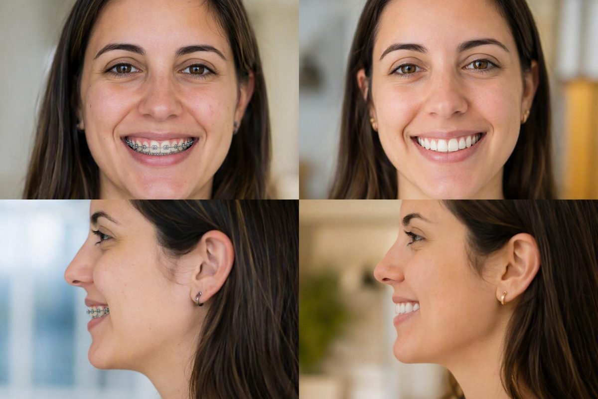
Searching for adult braces before-and-after photos is a reasonable way to explore what orthodontic treatment might change. A dramatic transformation may be genuine while still being a poor comparison for your teeth.
The available evidence also has an important limitation: the supplied galleries do not provide a standardized series of confirmed adult fixed-braces cases. Only one case is explicitly identified as an adult treated with braces. This article is therefore a guide to evaluating adult braces photos—not a representative adult-results gallery.
Adult braces may improve crowding, rotations, spacing, arch alignment, and some dental bite problems. Whether a particular change is feasible depends on the diagnosis, oral health, treatment mechanics, and individual goals. Even a closely matched case shows a possibility, not a promised result.
What adult braces before-and-after photos can—and cannot—tell you
A useful photo series may illustrate visible changes such as:
- Reduced overlap among crowded teeth
- Straighter-looking upper and lower arches
- Closed or redistributed spaces
- Improved tooth rotations
- A more even smile line
- Changes in how the upper and lower teeth appear to meet
- Differences in the visible dental midlines
Photographs are incomplete clinical records.
The most useful opening question is therefore not simply, “Do I like the after photo?” Ask, “What diagnosis and treatment produced the change between these photographs?”
Look for a fully labeled case
A credible adult comparison should disclose as many of the following details as possible:
- Age or age range: Was the patient an adult when treatment began?
- Diagnosis: Was the main concern crowding, spacing, an open bite, an underbite, a crossbite, or a combination?
- Dental versus skeletal involvement: Was the discrepancy mainly caused by tooth position, jaw position, or both?
- Appliance: Were fixed metal, ceramic, or lingual braces used? Were clear aligners involved?
- Extractions: Were teeth removed as part of the plan?
- Additional mechanics: Did treatment include elastics, expansion, temporary anchorage devices, or another appliance?
- Other procedures: Were restorations, implants, veneers, crowns, gum procedures, or jaw surgery part of the transformation?
- Duration: How long did active treatment last?
- Retention: Was a removable retainer, bonded retainer, or combination prescribed?
- Follow-up: Was the final image taken immediately after treatment or later?
Missing labels can change the meaning of a gallery. One treating-practice gallery, for example, combines braces and Invisalign results but does not identify the appliance or patient age for every image. It illustrates selected orthodontic possibilities but cannot confirm adult fixed-braces outcomes case by case (see the mixed-treatment gallery).
Check whether the photographs are comparable
Before judging the transformation, compare the technical quality of the images:
- Are the before and after views taken from the same angle?
- Is the head held in approximately the same position?
- Are lighting, magnification, and crop similar?
- Is the patient smiling equally in both images?
- Are the teeth together in the same bite position?
- Is one image brighter, closer, or more flattering?
- Did whitening or restorative work change the overall appearance?
- Did makeup, expression, or camera position affect the apparent facial change?
A single smile photograph is rarely enough. A more informative set includes a frontal facial smile, facial profile, upper arch, lower arch, and teeth-together intraoral views from the front and both sides. Arch views may reveal spacing and rotations hidden from the front, while side views help show changes in how the teeth meet.
Provider galleries are curated collections of selected cases. They do not establish a practice’s typical result, the frequency of complications, or what will happen to another patient. Unless the gallery supplies standardized records and follow-up, it should not be treated as evidence that the pictured result is common or durable.
What changes braces may produce, organized by the starting problem
The most useful way to interpret a transformation is by diagnosis rather than by how dramatic the smile appears. Two adults who both describe their teeth as “crooked” may have different space limitations, bite relationships, gum conditions, restorations, and jaw structures. Their appropriate treatment plans and finished results may therefore differ.
Crowding and tooth rotations
Crowding occurs when teeth do not fit neatly within the available arch space. Teeth may overlap, rotate, sit inward or outward, or become partly blocked from view.
After treatment, photographs may show:
- Less overlap
- Previously rotated teeth facing more evenly forward
- A smoother arch shape
- More consistent spacing between teeth
- Better alignment of teeth displaced toward the tongue or cheeks
The method used to create room is case-specific. An online non-extraction result does not prove that avoiding extractions is appropriate for another patient.
Photographs also do not show the complete relationship between the visible crowns and the roots beneath the gums. Nor do they establish how the supporting tissues responded around previously crowded teeth.
Gaps and generalized spacing
Orthodontic treatment may close spaces, redistribute them, or preserve them deliberately.
For example, a missing-tooth space might be:
- Maintained or reshaped for a future implant, bridge, or other restoration; or
- Closed by moving neighboring natural teeth when the diagnosis and mechanics make that suitable.
“All gaps closed” is therefore not automatically the best result. Symmetrical, intentionally preserved space may reflect a coordinated orthodontic-restorative plan rather than unfinished treatment.
Overbite
Overbite is the vertical overlap of the upper and lower front teeth. When that overlap is excessive, it may be described as a deep or excessive overbite.
The potential correction depends partly on its cause. A discrepancy driven mainly by tooth position may permit a substantial dental change. If jaw position is an important component, the treatment limits and goals may be different.
A full-face smile is not enough to assess an overbite. Teeth-together frontal and side views are more informative.
Underbite
In an underbite, the lower front teeth sit in front of the upper front teeth. Some adult underbites have a substantial dental component and may be improved through tooth movement. Others primarily reflect the relative positions of the upper and lower jaws.
That distinction matters because adult jaw growth is complete. Severe skeletal underbites may not be correctable with braces alone and can warrant an orthodontic-surgical consultation. Provider-authored guidance organized by bite type similarly distinguishes dental tooth-position problems from skeletal jaw discrepancies (review the bite-type guide).
Crossbite
A crossbite occurs when one or more upper teeth sit behind the corresponding lower teeth instead of outside them in the usual relationship. It may affect the front teeth, back teeth, one tooth, a larger dental segment, jaw width, or a combination of these features.
An after photograph may show the involved teeth in a different relationship, but a frontal smile can hide a posterior crossbite. Side and arch views are needed to understand what changed.
Open bite
In an open bite, some upper and lower teeth do not meet when the mouth is closed. A front open bite usually appears as a vertical space between the incisors even when the back teeth are together.
The potential result depends on whether the open bite is predominantly dental, skeletal, or mixed. Contributing tongue patterns or persistent habits may also matter to stability, so moving the teeth may not address every part of the long-term problem.
Dental midline discrepancies
The upper dental midline is the center line between the two upper front teeth; the lower midline is the corresponding line between the lower front teeth. These lines may not coincide with each other or with the center of the face.
Treatment may improve a midline discrepancy, but a perfectly centered-looking smile is not the only measure of success. The plan must also account for the bite, available space, tooth proportions, missing teeth, and relative positions of the arches. A small residual difference may be accepted in one treatment plan while a similar-looking difference is addressed in another.
Not every visible asymmetry requires treatment. Orthodontic care should respond to an individualized diagnosis and agreed goals rather than an assumption that every irregularity visible in a photograph is unhealthy.
A documented adult case: missing-tooth spaces closed over 30 months
The evidence pack contains one explicitly identified adult fixed-braces example. It is a treating-practice report, not an independently verified case study or evidence of a typical result.
The practice identifies the patient as a 27-year-old woman with spaces associated with multiple missing teeth. It reports using braces and temporary anchorage devices to move posterior teeth forward and close those spaces over 30 months (view the provider-reported case).
Its use depends on the movement required and the patient’s condition; it is not a routine requirement for every braces case.
Case summary
Field Provider-reported details Age 27 (provider gallery) Diagnosis Spaces associated with multiple missing teeth Appliance Braces Adjunct Temporary anchorage devices Duration 30 months Claimed result Posterior teeth moved forward and missing-tooth spaces closed Evidence limitations No independent verification, objective before-and-after measurements, standardized image assessment, complication reporting, retention follow-up, or evidence that another patient would qualify for the same plan
The case illustrates one possible alternative to preserving several spaces for later tooth replacement. It does not establish that every missing-tooth space can or should be closed.
The practice also reports an estimated saving from avoiding implants and crowns. That estimate should not be generalized: it was not independently verified, and both the cost and clinical suitability of orthodontic and restorative alternatives vary by case.
A reader with a visually similar gap may have different root positions, bone support, gum conditions, tooth sizes, restorations, or bite relationships. Those differences could favor preserving the space, closing it, or pursuing another plan entirely.
When changes become visible and why full treatment takes longer
Early movement and completed treatment are different milestones. Crowded teeth may begin to look straighter, or a small gap may narrow, before the upper and lower arches have been coordinated and the finishing objectives have been completed.
A cautious visual progression is:
- Early weeks: Movement may begin without being obvious in casual photographs.
- Around three to six months: Alignment or gap closure may become easier to see.
- Later months: Larger changes in crowding, arch coordination, or bite relationships may become apparent.
- Finishing phase: Treatment may continue after the smile looks straight while remaining details are addressed.
- Retention: Active movement ends and the plan for maintaining the result begins.
Provider-published guidance commonly places the first noticeable changes around three to six months and uses approximately 12 to 24 months as a broad complete-treatment benchmark. These are commercial-provider planning ranges, not independently established averages or promises for an individual patient (see the provider timeline overview).
Selected cases in the treating-practice gallery discussed above span approximately 12 to 30 months, but they involve different diagnoses, ages, appliances, and treatment plans. A limited 12-month treatment and a 30-month missing-tooth-space closure are not comparable predictive categories. The range demonstrates variability rather than a normal duration.
Treatment may continue after the front teeth look straight because visible alignment is only one possible objective. Depending on the plan, work may remain on bite relationships, space distribution, arch coordination, or preparation for restorative treatment.
Factors that may affect duration include:
- The type and complexity of the starting problem
- Whether the discrepancy is dental, skeletal, or mixed
- Gum, bone, and general oral condition
- Untreated decay or unstable dental work
- Missing teeth, implants, crowns, or substantial tooth wear
- Coordination with restorative treatment or surgery
- Missed appointments
- Broken brackets or other appliance problems
- Use of prescribed elastics or removable appliances
- Changes to the agreed treatment goals
Braces adjustments are often described as occurring about every four to six weeks, although the actual monitoring interval is individualized (review the provider’s adult treatment stages).
Some provider articles suggest adult teeth may sometimes move more slowly than teenagers’ teeth, but the supplied evidence does not quantify a dependable age-based difference. It is more useful to ask how your diagnosis, oral health, mechanics, attendance, and adherence affect your estimate than to assume adulthood automatically adds a fixed number of months.
Adult-specific planning: gums, crowns, implants, decay, and missing teeth
Adult orthodontic planning often involves more than arranging the front teeth. Assessment may need to consider:
- Gum condition and supporting tissues
- Existing bone loss or previous periodontal treatment
- Tooth wear
- Jaw function and comfort
- Available space
- Upper-to-lower bite relationships
- Fillings, crowns, bridges, or veneers
- Dental implants
- Missing teeth and future restorative plans
Active gum disease, untreated decay, or unstable restorations may need to be managed before active tooth movement begins. A history of dental treatment does not automatically prevent braces, but the plan must account for the current condition of the teeth, gums, and restorations. Provider guidance on adult planning specifically identifies gum condition, previous dental work, tooth wear, available space, and jaw function as relevant considerations (see the provider’s adult-planning discussion).
Crowns and fillings must be evaluated as part of the movement plan. If restorative work was completed after braces, the final photograph may not represent orthodontic treatment alone.
Dental implants require particular planning because they do not move orthodontically like natural teeth. When an implant is being considered but has not yet been placed, the orthodontist and restorative dentist may need to coordinate treatment order.
A missing-tooth space may be:
- Preserved for an implant, bridge, or another restoration
- Reshaped to provide the planned restorative dimensions
- Temporarily occupied by an artificial tooth during treatment
- Closed through orthodontic movement in a selected case
The reported 27-year-old TAD case illustrates the final pathway but does not prove it is suitable for everyone. Feasibility depends on the location and number of missing teeth, surrounding tooth positions, bite, bone and gum condition, and overall treatment goals.
A visually similar online case cannot answer those questions. An individualized examination and appropriate imaging are needed before concluding that a tooth can be moved, a space can be closed, or an existing implant can be incorporated into treatment.
Dental movement versus skeletal change—and what may happen to the face
“Will braces change my face?” is partly a question about what caused the original appearance.
A dental discrepancy is driven mainly by tooth position. Front teeth may, for example, tilt forward or backward and affect apparent lip support. A skeletal discrepancy involves the relative positions or proportions of the jaws. Many patients have a combination of the two.
Adults have completed jaw growth, so childhood-style growth modification should not be assumed. Braces can move teeth and may improve some dental components of a bite problem, but they do not make an adult jaw grow predictably in a new direction.
Severe skeletal underbites, open bites, or other major jaw discrepancies may require an orthodontic-surgical consultation. If an online transformation involved jaw surgery, braces were one part of combined treatment; the full facial or jawline change should not be attributed to brackets and wires alone.
Tooth movement may nevertheless alter appearance by changing apparent lip support, smile display, or profile balance. Such changes may be more noticeable when the starting dental discrepancy is substantial. Provider guidance characterizes facial changes in adults as generally subtler than those possible during growth and states that braces do not physically enlarge the lips (see the provider’s facial-change discussion).
Be skeptical of promises that braces will reliably:
- Produce dramatic adult jaw growth
- Create a universally sharper jawline
- Tighten facial skin
- Remove wrinkles
- Make every patient look younger
- Reproduce the facial result shown in a selected case
A responsible prediction should distinguish changes expected from tooth movement from those that would require surgery or restorative care. It should also identify the aspects of facial appearance that cannot be promised.
Fixed braces versus clear aligners: compare the plan, not just the final photo
A finished photograph may not reveal whether the patient used fixed braces, clear aligners, elastics, expansion, temporary anchorage devices, surgery, restorative treatment, or a sequence combining several methods.
Fixed braces remain attached to the teeth. They may provide useful control for some complex rotations, crowding, and bite movements. Their fixed design also removes the daily decision of whether to wear the appliance, although patients still need to maintain hygiene, attend appointments, follow instructions, and use elastics when prescribed.
Clear aligners are removable. That can make eating and cleaning routines more flexible, but treatment depends on following the prescribed wear schedule and managing the trays consistently.
Neither option is universally superior. Suitability depends on:
- The diagnosis and exact movements required
- The complexity of the bite correction
- Whether supporting mechanics are needed
- Visibility preferences
- Eating and cleaning routines
- Willingness to manage a removable appliance
- Appointment and monitoring requirements
- Ability to follow the complete plan
An unlabeled gallery mixing braces and aligners cannot show which appliance produced a particular result. It is especially unreliable when cases also differ by age, diagnosis, duration, and additional procedures.
Adults can use Aligner Source’s first-party comparison of metal, ceramic, self-ligating, lingual braces, and clear aligners to review practical differences in visibility and daily routine. It should not be treated as independent comparative clinical evidence.
Instead of asking which appliance produces the best-looking after photo, ask:
- Which movements does my treatment require?
- What limitations does each option have in my case?
- Would either plan require elastics or other adjuncts?
- How will progress be assessed?
- What happens if movement does not proceed as planned?
- Does the estimated duration cover active treatment only?
- What retention plan follows each option?
These questions compare actual treatment plans rather than marketing categories.
The after photo is not the endpoint: retention and consultation questions
Teeth can change position after active orthodontic treatment, which is why retention is part of the treatment plan rather than an optional cosmetic extra.
Possible approaches include:
- A removable retainer
- A fixed or bonded retainer
- A combination of fixed and removable retention
No single retainer type or wear schedule is right for everyone.
Retention may not be visible in a frontal smile. One supplied treating-practice gallery caption identifies a bonded retainer, illustrating that it may only be apparent in an appropriate intraoral view (view the labeled retention example).
A photograph taken immediately after appliance removal cannot establish long-term stability. Ask when the image was taken, what retention was prescribed, and whether later records exist.
Questions to ask during your consultation
- What is my complete diagnosis?
- Is the discrepancy primarily dental, skeletal, or both?
- Which teeth are planned to move, and in what direction?
- What are the goals for alignment, spacing, and bite?
- What limitations or compromises should I understand?
- Will I need elastics, extractions, expansion, or temporary anchorage devices?
- Could restorative work or gum treatment be required before, during, or after braces?
- Is surgery a possibility in this plan?
- What is included in the quoted fee?
- Are imaging, emergency visits, replacement appliances, and retainers included?
- What duration range applies to my diagnosis?
- What circumstances could extend treatment?
- What retainer is planned?
- How will the result be monitored after active treatment?
Questions to ask about a “comparable” case
- Was the patient an adult at the start of treatment?
- What was the formal diagnosis?
- Was the problem dental, skeletal, or both?
- Which appliance was used?
- How long did active treatment last?
- Were teeth extracted?
- Were elastics, anchorage devices, restorations, or surgery involved?
- Were the images standardized?
- When was the after photo taken?
- Is later retention follow-up available?
- What important differences exist between that patient and me?
Frequently asked questions
How long do braces take for adults?
Provider-published estimates commonly use approximately 12 to 24 months as a broad adult active-treatment range, but this is not a guaranteed average. Limited movement may take less time, while complex bite correction, missing-tooth management, restorative coordination, or skeletal involvement may take longer.
Early changes may become visible around three to six months, but cosmetic improvement is not the same as completed treatment. The provider-reported missing-tooth case discussed above lasted 30 months.
Can adult braces change your face or jawline?
Braces can change tooth position, which may subtly affect apparent lip support, smile display, or profile. Adults should not expect braces to produce childhood-style jaw growth.
If facial imbalance is caused mainly by a severe skeletal discrepancy, braces alone may be insufficient and an orthodontic-surgical consultation may be appropriate. Braces do not physically enlarge the lips or guarantee a sharper jawline.
Can braces close gaps caused by missing teeth?
Sometimes. A missing-tooth space may be closed by moving neighboring natural teeth, or it may be preserved and shaped for a future restoration. The appropriate choice depends on the space’s location, surrounding teeth, bite, gum and bone condition, and restorative plan.
Temporary anchorage devices can assist selected movements, as illustrated by the treating-practice case involving a 27-year-old patient. That example does not establish that every missing-tooth gap can or should be closed.
Can teeth shift again after braces are removed?
Yes. Teeth can change position after active treatment, so a retention plan is normally used. Depending on the case, it may involve a removable retainer, a fixed retainer, or both.
Long-term stability cannot be judged from a photograph taken immediately after braces are removed. Ask what retention was prescribed, whether later follow-up exists, and when the final image was captured.
How can I tell whether a before-and-after case is comparable to mine?
Start with the diagnosis rather than visual resemblance. Confirm the patient’s age, the type and severity of the problem, appliance, duration, extraction status, adjuncts, restorative work, surgery, and retention plan.
The photographs should also use consistent views, lighting, magnification, head position, expression, and bite position. Even when those details match, the case remains an illustration rather than a prediction because gum condition, roots, bone support, restorations, jaw relationships, and treatment goals still require individual assessment.
The ideal after result is more than visibly straight front teeth. It should reflect the individualized goals established for alignment, bite, oral health, function, comfort, and stability.
Use online adult braces before-and-after cases to prepare informed questions—not to diagnose yourself or predict an identical transformation. Ask an orthodontic professional for fully labeled adult cases relevant to your diagnosis and for a personalized active-treatment and retention plan. Aligner Source provides general educational information, and its terms direct individual treatment decisions to the reader’s orthodontist (read the informational-use notice).